From Ultra-Wide Field TrueColor, Autofluorescence, and fluoresceine angiography, iCare EIDON Family is ready for enhanced diagnostics of eye diseases, such as diabetic retinopathy, geographic atrophy, and AMD. Discover how to improve treatment and overcome the hurdles in your clinic or hospital environment.
Ophthalmology clinics are often buzzing, and imaging is the bottleneck. Nurses wheel in patients from neurology, diabetes, and post-op wards. Concurrently, the staff struggles with aging fundus cameras—manual alignment, re-focusing, constant retakes and dilation slow things further, and decisions can’t be made without clear and comprehensive visuals. With no room in the schedule, staff skip breaks, patients wait, and the routine becomes rushed. With the iCare EIDON Family fundus imaging device, there is an option.
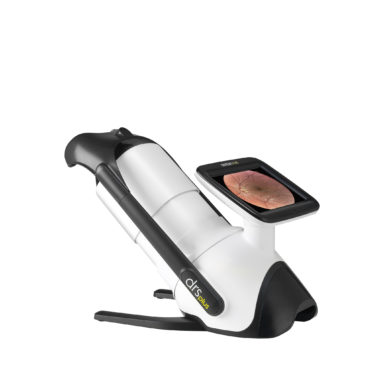
Why choose an iCare EIDON Family fundus imaging device
iCare EIDON Family is ideal for advanced retinal imaging, delivering high-resolution, Ultra-Wide flied, and True-Color confocal images. It’s a perfect fit for ophthalmology clinics, hospitals, and eye care professionals who require detailed diagnostics and superior image quality. The five main benefits of iCare EIDON Family are:
1. Examination of almost the entire retina with UWF Module
The early signs of many pathologies are often subtle and appear first on the retina’s periphery, which the standard field tests can’t reveal. Previously, only about 50% of the retina could be visualized, but the Ultra-Wide Field enables examination of almost the entire area—significantly enhancing the ability to detect diseases early. iCare EIDON Family provides widefield optics to allow the imaging of the central retina and the periphery, facilitating improved and early diagnosis. For example, the Ultra-Widefield Module captures 120˚ images of the retina in a single shot and even up to 200˚ with the Mosaic functionality.
2. Unparalleled image quality for quick and reliable image interpretation
Confocal imaging surpasses conventional fundus photography by blocking back-scattered light outside the retina’s focal plane, delivering sharper, higher-resolution, and higher-contrast images. As a result, you’ll get images that are easier to interpret and help to identify specific conditions, such as diabetic retinopathy, with much better visualization.
iCare EIDON combines a confocal optical engine with a white light LED source. The device illuminates the retina and captures TrueColor fundus images characterized by colors close to reality—the retina appears as it looks when directly observed, as the entire visible spectrum is present in the image. Blocking scattered light ensures clarity even with cataracts and works without dilation for pupils as small as 2.5 mm.
In addition to white LED, the iCare EIDON red-free filtering enhances the visualization of retinal vasculature, and blue images provide an improved view of the retinal nerve fiber layer (RNFL). Even if there is an obstruction in the eye that would typically block light, it still allows for precise imaging of the retina.
3. More comprehensive eye examinations with versatile imaging modalities
In addition to the standard model, iCare EIDON AF provides the added advantage of autofluorescence imaging, allowing the assessment of the integrity of the Retinal Pigment Epithelial (RPE) layer. The confocal scanner guarantees high details and contrasts autofluorescence images with high-resolution quality in one shot without the need for image averaging. It also offers 120° field of view for single shot capture and up to 200° for mosaic mode. As with the standard model, any staff member can run the tests, speeding up the clinic workflow. Concurrently, the device enables a quick and comfortable patient experience.
Furthermore, iCare EIDON FA complements the range of imaging modalities with fluorescence angiography. The device also offers the advantage of quickly capturing a detailed high resolution FA video, allowing the user to focus on the patient. The recordings provide a realistic and dynamic view of retinal vasculature and circulation mechanisms that may be missed with static flash photography. The user can choose the best from multiple frames to document the pathology.
4. Add patient comfort and efficiency with dilation-free procedure
With the iCare EIDON Family fundus imaging devices, no pupil dilation is required. The patients don’t have to wait for the drops to take effect, wear sunglasses, and wait before driving afterwards—meaning quick and comfortable examinations, reduced appointment times, i.e., more satisfied customers, and improved workflow and efficiency.
5. Streamline your workflow through automation and human-centric design
iCare EIDON Family makes fundus imaging easy, comfortable and standardized. Using the devices requires no special training. The automation assists the users, many of whom are trainees. Instead of manual tuning, they can just push the button. Switching from manual to automatic operations regarding alignment, focus, exposure, capture, and mosaic is also possible. In addition, the ergonomic, motorized chin rest, soft flash and no need for dilation provide added comfort to patients.
As a result, you can speed up examination time and improve workflow.
Ready to upgrade your clinic with an easy-to-use, versatile, high-quality fundus imaging device?
Let’s make the world see.