Originally published by MiVision
Dr Malvika Gupta explains why the iCare EIDON Ultra-Widefield fundus camera is a “jack of all trades and master of many” – and why that makes it a necessity in practice, even if it’s not a billable item.
Eye care professionals are well aware of the need to visualize the peripheral retina when diagnosing and managing retinal disease. With the evolution of new technology, this is becoming easier to do and the details provided are increasingly revealing. Devices like iCare’s EIDON Ultra-Widefield, with TrueColor confocal technology, are also providing valuable opportunities to educate patients about their disease states, in the first instance and as time goes by.
For ophthalmologist Dr Malvika Gupta from Eastern Eye Specialists in Melbourne, having “fought to invest in the iCare EIDON for three years at the Sunbury practice” this device has become a clinical necessity that she wouldn’t be without.
“I’m a pioneer in wanting to provide good patient education and I knew that this device would enable me to do exactly that. It provides a platform from which you can literally share information you see with your patient – you can show them what you are talking about.
“And it’s also an extra safeguard. In busy retinal practices you can easily miss things you’re not looking for when you’re viewing a cataract, the optic disc, and the macula, so having a fundus camera is a safeguard against that – even a tiny spot is visualised under magnification – right there in your face,” she told mivision.
Getting from A to B
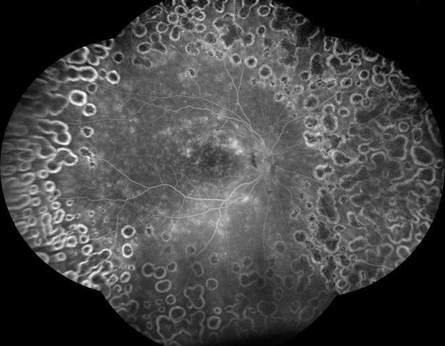
With an expanded field of view of the retina up to 200°, iCare’s Ultra-Widefield imaging technology provides more information about the signs of pathologies in the periphery. Besides the TrueColor confocal images, it enables the acquisition of Ultra-Widefield infrared, autofluorescence, and fluorescein angiography images.
Dr Gupta said many eye care professionals will be hesitant to invest in the device because “it is not a billable item” and therefore there is “no financial or economic reason to have the device”. However, for her, the benefits it delivers far outweigh the costs.
“It’s like buying a car. Any car will get you from A to B, so buying a luxury car is not a necessity – you don’t have to pay for one, but having invested in one will bring in more ease and make the experience more enjoyable and worthwhile.
“As ophthalmologists, we spend two-thirds of our lives at work, we’re workaholics, so why not pay more attention to the experience and spend a little more on the devices that enhance our clinical experience and the experience for our patients.”
Encouraging compliance
Dr Gupta said the device can be invaluable when getting patients onboard with their recommended treatment plan. Using the Eidon’s images, she can now show patients the true picture, literally and then over time, demonstrate the change being achieved.
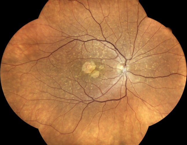
“The images are crystal clear, with perfect contrast, and a high degree of peripheral visualisation – it’s easy to show the normal and the abnormal, which makes it easy for patients to understand complex messages,” she said.
When a patient is being treated with anti-VEGF for a macular hemorrhage for instance, she shows them, over time, how it disappears following injection therapy. “Otherwise, they come in, have an injection and they don’t see what’s going on. It is very rewarding and includes patients in their own care.
“Similarly, I can show them a scar and they can see what it looks like. When a patient’s life is going to change based on a diagnosis, their ability to see it and come to terms with it visually is comforting for them and satisfying for me as a clinician.
“So, I feel it’s a necessity – there’s comfort in sharing complex clinical information with the patient and them being able to have that same realisation as I do.”
Of greatest value is explaining eye disease to patients who have few symptoms.
“There are patients with poor vision, and they will be motivated to come on board with treatment to recover what vision they’ve lost, or at least maintain the vision they have. “The more challenging patients are those who have reasonably good vision – and this is where the motivation is important.
“Someone with 6/7.5 vision, who is not particularly symptomatic, has their diabetes reasonably under control, yet may be developing worsening eye disease. I pick up early signs but because they’re not noticing any change, they can’t comprehend why I want them to see me more regularly. It’s hard for them to fathom the reality of disease severity and progressive nature.
“Communication flows easily when you can highlight their own pathology and compare it with another image, showing normal or far advanced disease,”she explained.
“With the camera, I can show a patient their leaking microaneurysms and proximity of the exudates to the foveal centre. This makes the threat real and motivates them to manage their systemic diseases, like diabetes and hypertension, better and be more compliant to frequent visits. They will come back every three months and, with fundus imaging, together we can monitor the change and manage as needed.”
Dr Gupta said it’s also helpful when you want to reassure a patient that treatment is not necessary.
“I had a patient with a microaneurysm. They often rupture and spontaneously heal but if leaking into the macula persists, treatment is probably needed.
“Rather than telling the patient, I was able to show them that the microaneurysm was located away from the fovea, with no immediate threat of leakage into the macular centre or line of sight. This helped explain why I didn’t feel the need to treat them. That was immediately reassuring for the patient. Sharing imagery of the disease signs helps justify a treatment plan so much more effectively than a one-sided didactic conversation.”
Connected devices
Having had the iCare EIDON Ultra-Widefield for a year now, Dr Gupta said it has been a worthwhile investment and, despite a few teething issues, a “set and forget”.
“There have been a few teething issues – something as simple as patients wearing masks while doing the scans caused the lenses to fog. We were getting really poor scans and we couldn’t figure out why. The Designs for Vision) rep suggested the patients take their masks off and life changed.
The machine did not know that (mask wearing) would ever be a challenge, and we were not aware of the possibility.
“Another issue that took us by surprise was the impact of lighting – we had to place the camera in a brightly lit area, and we couldn’t dim the lights enough to enable good images. The rep suggested a veil over the device when taking the pictures, which worked brilliantly. So, you have to provide the right circumstances for the camera to be able to function.
“We’re amazed by the picture quality, we make montages all the time – it’s beautiful – you’re taking the pictures, auto fluorescein images, angiograms, and creating montages, all from one device, which makes it more useful.
“Some might say that the iCare EIDON must be a ‘jack of all trades, master of none’ but I don’t believe in that – if you want to specialise in one thing you can, but is that what’s really needed; what you will actually use day-to-day? Just because this device does five things, that does not mean it’s not going to be good enough. I’d suggest those people with doubts try it. I think it is ‘jack of all trades – and master of many!”
With cloud connectivity and devices increasingly integrated from clinic to operating theatre, Dr Gupta said she strongly feels the equipment and diagnostics she invests in must have the potential to talk to each other – if not now, then in the near future.
With this in mind, she’s decided that it makes sense to “keep it in house” by buying from one supplier who will be able to assist with upgrades, software, and the integration of devices into one platform.