Early and reliable detection of retinal diseases like diabetic retinopathy, geographic atrophy and AMD is crucial. However, traditional fundus imaging poses challenges that escalate in overcrowded healthcare settings, and at small clinics and optician shops without sufficient expertise. Discover how the iCare fundus imaging devices overcome hurdles and benefits your daily operations.
Using the iCare fundus imaging device, even trainees working in busy hospital environments can perform eye examinations reliably. Also, small clinics and optician shops can provide more comprehensive services for their customers, while improving workflow, efficiency, and patient satisfaction. Today, more and more patients have their fundus images taken efficiently with the iCare fundus imaging devices, leading to earlier detection and intervention.
How to interpret fundus images more reliably
Detecting subtle retinal abnormalities can be difficult even for experienced professionals. Technically, the challenge stems from the image quality and coverage. Reproduced fundus images provided with traditional devices often appear reddish or greenish, potentially obscuring crucial details. Even filters fail to compensate for the shortcomings.
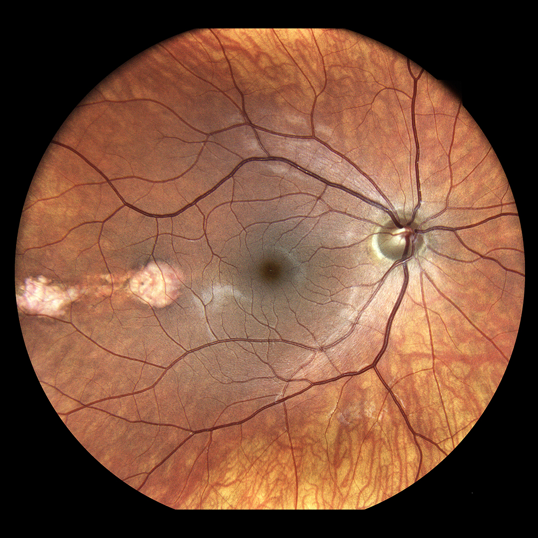
iCare revolutionizes fundus imaging by combining a confocal optical engine with white light LED. The device illuminates the retina and captures TrueColor fundus images characterized by colors close to reality. Confocal imaging surpasses conventional fundus photography by blocking back-scattered light from outside the retina’s focal plane, delivering sharper, higher-resolution, and higher-contrast images. It maintains quality even with media opacities like cataracts and functions with pupils as small as 2.5 mm without dilation.
With white LED, the retina appears as it looks when directly observed, as the entire visible spectrum is present in the image. In addition to white LED, red-free filtering enhances the visualization of retina vasculature, and blue images provide an improved view of the retinal nerve fiber layer (RNFL). The red channel allows light to penetrate the retina’s deep layers; in turn, infrared light provides detailed information corresponding to the choroid. As a result, ophthalmologists can identify specific conditions, such as diabetic retinopathy, with much better visualization.
Towards more comprehensive and detailed eye examinations
Early diagnosis can prevent eye diseases from progressing to a stage requiring treatment. The challenge is that early signs of many pathologies are often subtle and appear first on the retina’s periphery, which the standard field tests can’t reveal. Previously, only about 50% of the retina could be visualized.
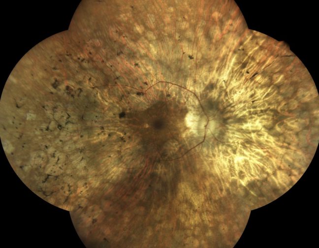
Unlike traditional devices that capture limited retinal areas, iCare’s widefield optics provide a detailed view of the central and peripheral retina, allowing earlier and more accurate diagnoses of conditions like diabetic retinopathy. Autofluorescence imaging enables detailed assessment of the Retinal Pigment Epithelium (RPE) layer, providing ultra-high-resolution images in a single shot.
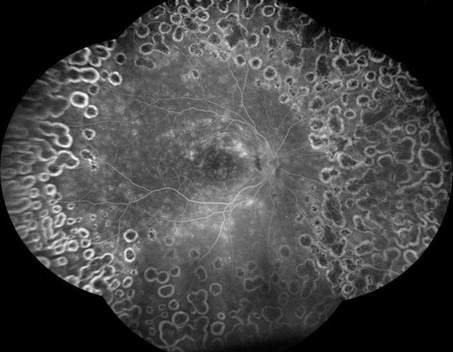
In turn, confocal technology enhances imaging clarity by penetrating media opacities, while widefield autofluorescence expands retinal visualization up to 160°. Based on these innovations, Fluorescein Angiography (FA) offers high-resolution video capture for a dynamic, real-time view of retinal vasculature, improving pathology documentation.
Furthermore, to enhance early disease detection, iCare has introduced Ultra-Widefield Imaging, capturing up to 120° in a single shot and 200° with the Mosaic function. The module allows for comprehensive retinal imaging across TrueColor Confocal, Infrared, Autofluorescence, and FA modalities, significantly improving diagnostic capabilities.
Making fundus imaging faster and more patient-friendly
Manual fundus imaging requires expertise due to the complexity of capturing high-quality retinal images. Alignment and focus demand precision, as the slightest misalignment could compromise image clarity. Technicians must also carefully adjust light exposure to prevent overexposure or underexposure, which could obscure crucial retinal details. Thus, image quality varies significantly depending on the operator’s skill.
The process also demands a dimly lit examination area, further complicating workflow. Concurrently, traditional imaging requires pupil dilation. The patients must wait for the drops to take effect, and it’s difficult to capture sharp images if patients move. Afterwards, they must wear sunglasses and wait longer before driving after the procedure. All this adds time to appointments and creates more congestion in busy clinical settings.
Using iCare’s automated fundus imaging, you can just push the button and have a perfect image quickly and comfortably without dilation. The device allows switching from manual to automatic operations regarding alignment, focus, exposure, capture and mosaic. The ergonomic, motorized chin rest, soft flash and no need for dilation provide added comfort to patients.
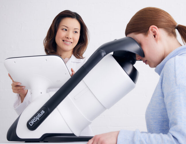
The tools of the trade: iCare EIDON and DRSplus
Both iCare EIDON and DRSplus deliver high-quality fundus images, with iCare EIDON being the more versatile option for advanced diagnostics. iCare DRSplus was created as a lighter alternative to iCare EIDON, addressing the needs of smaller clinics that do not require comprehensive diagnostic imaging.
iCare EIDON is ideal for hospitals and specialized clinics, which can serve as a primary diagnostic tool or be used as part of initial screening workflow before referring patients to university hospitals for further examination, iCare DRSplus is sufficient for standard fundus imaging, covering the standard field of view. It is best suited for optical stores, general practitioners, small clinics, and even in non-ophthalmic setting, such as diabetes clinics, that do not require advanced diagnostic capabilities—an excellent choice for being implemented as a part of preliminary screening workflows. Beyond imaging, the iCare DSRplus integrates intelligent software to enhance diagnostic accuracy. AI-powered analysis assists in detecting abnormalities, helping healthcare providers make more informed decisions faster. This not only improves patient care but also streamlines workflow in busy practices.
The choice is yours.